
I know what you're thinking: "red light therapy for eye health and vision, does that even work!?"
It turns out that the scientific evidence on red light therapy for vision and eye health is pretty impressive. Whether you're talking about different eye condtions such as glaucoma, age-related macular degeneration, or myopia, the results give lots of hope.
I went through all 200+ studies on red light therapy for eye health and analyzed the outcomes for you. The full results are listed below in this blog post. If you don't have the time to read all my findings, read my summary below.
For the best all-round results, you'll want the light to enter your eyes directly. Red light has the best evidence base, although near-infrared has many positive studies in its use as well.
You'll probably want to use near-infrared for eye health as well to reach deeper tissues of the eye, such as the retina and macula.
For the best results, you'll have to expose your eyes to 40 - 60 mW/cm2 for up to 3 minutes per day. You can reach these results by standing 10-12 inches from many modern panels, keeping your eyes open, and then wearing protective glasses when you are done.
From a safety perspective, please keep in mind that some studies use 100 mW/cm2 per second or more, but I'm not going to recommend that as the 40 - 60 mW/cm2 range for 3 minutes is simple to use and gives good all-round results.
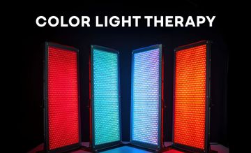
If you want a cheaper treatment option, get a tabletop panel or even a handheld panel and use that for 3 minutes of treatment at my suggested power output.
For the best results you may want a combination of 630 nm, 660/670 nm, and a near-infrared wavelength, although most studies use either alone. Hence, more comparison studies are needed to compare different red wavelengths with each other, and compare red against near-infrared.
And just to be clear: the light needs to reach your eyes and penetrate them, such as through the lens and pupil, to reach the retina, macula, optic nerve, and other tissues. So if you wear protective glasses all the time, you won't get the full benefits, although you will get benefits from systemic effects as treating the brain affects the eye area also.
Generally, if you follow the common-sense treatment guidelines that I've posted above, there shouldn't be any side effects. If you exceed the guidelines, you can damage your eyes, such as retinal damage. Almost all studies don't show any side effects though!
To fully avoid any side effects, I still recommend seeking out professional medical help - so nothing in this blog post is medical advice. Also, if you've got surgery for your eyes, I'm not willing to take the risk of giving any recommendations here.
In the full blog post, I walk you through the basics of eye anatomy, all the research on red light therapy for the eye conditions that exists right now, why you should be focusing on overall health and not just red light therapy for eye health, and much more.
Continue reading below if you want to know the full story!
Update: Interview
If you wish to view an interview I did about this topic, check YouTube here:
Eye Anatomy Basics
First up, I'll say a thing or two about eye health and eye anatomy. You'll need to understand these to later understand why red light therapy for the eyes works or not and how it works.
The eyes are integral to the human experience (1; 2). About 2.5 millennia ago, Aristotle already stated that humans derive a certain pleasure from using our sensory organs, especially the eyes. Nowadays, it turns out that 50% 0f our sensory receptors are located in those eyes (2).
The eyes apprehend visible light - all the colors of the rainbow. Other light such as infrared and ultraviolet affect the eyes as well, but are invisible perceptually. The eye allows you to see the world around you - but has other functions as well such as keeping time. Timekeeping, here, entails the roughly 24-7 circadian rhythm that not only affects your eye but your brain and entire physiology (3; 4; 5).
Put in plain English, the physiology of your entire body responds through the light that enters the eye, or the absence of light. That eye is directly connected to the brain with the optic nerve. And, the brain has an area called the "suprachiasmatic nucleus" that acts as the central clock of your body (6; 7; 8).
The rest of the body (ideally) follows that central clock and as a result, physiology works optimally. Light and darkness are like ebb and flow and both necessary for optimal eye health and overall bodily health (9; 10; 11).
So let's consider the basic structures that make up the human eye:
Here, you can see the basic structures of the human eye that are visible from the exterior. These are:
- The sclera - the white part that's visible in the eye (12; 13; 14). The sclera isn't just a passive structure - it helps provide stability to the other parts of the eye. The sclera is made up of collagen and surrounds the entire eye, not just the front. The sclera can also be affected in eye disease, such as myopia.
- The black pupil - which allows for the entrance of light into the eye (19; 20; 21; 22). The pupil has more functions though, and will change size based on arousal, wakefulness, and is affected by human emotion!
- The green-yellow colored iris - the iris can increase or decrease in size, and therefore control how much light enters the pupil (15; 16; 17; 18). Melanin in the eye co-determines your eye color. And eye color can affect how the eye functions, such as in glare situations. The iris can also be affected in eye diseases.
- The cornea is placed over the pulis and iris. The cornea is technically invisible in the picture above. The sclera are not covered by the cornea. The cornea can be affected by eye disease, such as dry eyes (19; 20; 21; 22; 23). The cornea is also very metabolically active and has many nerve endings. Infection, chemicals, or mechanical trauma can all affect the cornea and therefore overall eye health. Overall eye health can also affect the cornea. You can envision the cornea as a protective layer for the pupil and iris.
- The eyelids above the eye - these help you reduce light input and are closed when you're resting or asleep. So the eyelids can prevent light from reaching the iris and pupil. The eyelids aren't just passive structures though, they play a fundamental role in blinking and in keeping your eyes hydrated and even nourished (24; 25; 26; 27). Just think about it, people who stare and don't blink eventually end up with dry eyes. The eyelids also protect against mechanical and chemical trauma. In some neurological and eye diseases, the eyelids can also be excessively closed.
Next up, we've got the following anatomy of the back of the eye, so the structures that are found behind the pupil, iris, and sclera:

I won't consider all structures in the picture above but just the most important ones:
- Lens - this structure allows you to focus your eyesight, thereby allowing the eye to focus on objects nearby you or far away (28; 29; 30; 31). The lens can be affected by disease - that's why in part you may need glasses or artificial lenses. The lens can also be affected by cataracts and other conditions. In cataracts, your lens becomes cloudy and you won't be able to see like normal. As you can imagine, other problems can also occur with the lens, such as displacement which makes vision more problematic.
- Retina - the backend of the eye where the light that travels through the pupil and lens ends up at (32; 33; 34; 35; 36; 37). The physiology and makeup of the retine is extremely complex and even I didn't study that topic in great detail. Nevertheless the retina can be affected by many different diseases - some acquired from birth and others in time. The retina is very metabolically active and affected by lifestyle diseases, such as hypertension and diabetes type II. Retinal blood flow is also very high. Diet and exercise greatly impact retinal health - a topic I'll cover later in this blog post.
- Macula - the inner central part of the retina that's most specialized in the clarity of vision (38; 39; 40; 41; 42). The macula can degenerate due to age (Age-related Macular Degeneration (AMD)) or through other factors. Myopia is also often caused by the macula, in part. Diet is also, once again, extremely important to macular health. Chemical exposure can also affect macular health.
- Choroid - is a blood supply area that's located between the retina and the white of your eye that I discussed earlier, the sclera (43; 44; 45; 46). The choroid can also change its thickness, therefore moving the retina and affecting your vision. The choroid can be affected in many different eye conditions, such as tumors, myopia, AMD that I talked about earlier, as a side effect of diabetes, and more.
- Optic nerve - this nerve runs from the eye directly to the brain (47; 48; 49; 50; 51). You can simply see the optic nerve as a transmitter, that relays the light/data that enters the cornea, pupil, lens, retina, macula, etc, and transfers it to the brain. With a dysfunctional optical nerve, you can imagine that vision becomes harder - and that's precisely what can be the problem in neurodegenerative conditions such as Multiple Sclerosis. Many other diseases can affect optic nerve function, however, as well as aging.
I hope I've explained the most basic structures of the human eye in simple terms. Don't worry too much if you cannot fully remember these structures. Later on in this blog post, I'll state the benefits and risks of red light therapy for eye diseases in terms that are as simple as possible.
Next up, let's consider a bit more about why eye health matters so much:
Eye Health Basics
Eye health doesn't just affect your eyes. The eyes are not just the "mirror of your soul" - as the popular saying goes - but are also a very accurate reflection of overall human health (52; 53; 54; 55; 56).
Eye health, for instance, is related to your quality of life (52; 53). Poor eye health is also associated with increased morbidity and mortality (52). Eye problems are also widespread, with 1 billion people worldwide either having an impairment or full-fledged blindness (53). Other estimates are different, assuming that dry eye disease alone affect 1 in 11 people worldwide (56).
Here's what a recent review states about eye disease:
"Incidence of cataract, diabetic retinopathy, macular degeneration, and glaucoma will significantly increase by 2050. Visual impairment can increase morbidity and mortality in nonocular disease. There are different patterns of vision loss in cataract, diabetic retinopathy, age-related macular degeneration, and glaucoma." (55).
So, the eye disease problem is poised to become worse, if things continue as they are.
Fixing these problems, in turn, improved quality of life. And, fixing eye health problems also has economic benefits for individuals and society at large (54). Also, consider a few of the associations between eye disease and other health conditions:
- Eye conditions increase demetia risk by 20-30% (57). Dementia, such as Alzheimer's, also increases eye disease risk (58). And you can even measure risk of Alzheimer's by biomarkers in the eye (59).
- Eye conditions and heart and blood vessel health are intertwined (60; 61; 62). If you do worse on heart disease risk factors it's likely you'll do worse in terms of eye health as well. One reason here is that the eye functioning and health is highly dependent on blood flow.
- Diabetes and eye disease risk are perhaps most famous and also interrelated (63; 64; 65).
- Eye disease is even one of the characteristic symptoms of early Parkinson's Disease (66; 67; 68).
- Kindey disease is intertwined with poorer eyesight, and vice versa (69; 70; 71; 72; 73). Here, you can monitor changes in the eye such as the blood vessel and therefore infer greater kidney disease risk.
Controlling for many of these risk factors for diseases such as heart disease and diabetes, in turn, also reduces the risk of eye diseases. Let's find out:
- Blood pressure increases the risk of many different eye diseases (74; 75; 76).
- Insulin resistance increases the risk for many eye diseases (77; 78; 79; 80).
- The body fat percentage, in particular being overweight or obese increases eye disease risk (81; 82; 83; 84).
- Waist circumference is even more strongly linked to eye disease than just BMI or being overweight (85; 86; 87; 88). So in many cases, if you want to improve eye health or decrease the risk of eye disease, the simple but uncomfortable truth is to normalize your body fat percentage. For every 5 centimeters of waist circumference increase (2 inches, give or take), your risk of cataracts and macular degeneration increases by 20% (89).
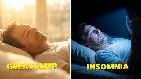
- Sleep quality is linked to eye health (99; 100; 101; 102; 103; 104; 105; 106; 107; 108; 109; 110; 111). Eye disease also often reduces sleep quality!
Why do I mention many of these risk factors above? Because they'll return later on. If you didn't know, red light therapy affects many of these risk factors that I've mentioned above. So, instead of focusing on the eyes only, if you consider the human body as a whole, red light therapy can help you improve these risk factors. If you want content on these topics, consider:
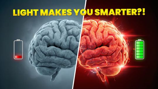
- Red Light Therapy For Upgrading Your Brain Health - for lowering Parkinson's Disease and Alzheimer's risk.
- Does Vielight Work? A Close Look At The Spectacular Vielight Science - a very useful device for treating the brain!
- Red Light Therapy For Weight Loss: The Science Of Supercharging Fat Loss - lose body fat to reduce eye disease risk. Your body fat percentage is also closely intertwined with blood pressure levels!
- Red Light Therapy For Sleep: The Science of Sweet Dreams - this blog post considers all of the studies on using red light therapy for better sleep quality.
- The Effectiveness Of Light Therapy For Sleep Disorders - this blog post considers red light therapy for sleep conditions - which you should fix if you can!
Next up, let's consider how red light therapy specifically and light therapy in general affect the eyes:
General Red Light Therapy For Eyes Benefits
Let's now consider some of the implications of red light therapy for eye health benefits. I'm using Vladimir Heiskanen's excellent database, which currently contains almost 8,000 studies.
We've got quite a few reviews published in the last few years on light therapy for eye health, in the 2020-2024 area (90; 91; 92; 93; 94; 95; 96; 97; 98). I've included these most recent reviews because they take the most recent evidence into account also. If you didn't know, a review in medical science analyzes and integrates previous studies into a new overview.
The good thing about these reviews is that the scientists creating them have spent hundreds of hours on these topics. And, often, these scientists have a specialized career in this field. Let's consider the most important lessons that I got from reading through these reviews:
- First up, it's not just red light therapy that's helpful for eye health, but the entire visible spectrum as well (90). Wavelengths from 500 nanometers to 1,000 nm are mentioned (90). I'll say that often even researchers aren't aware of the effects of wavelengths such as 1,064nm.
- Researchers also mention the value of LEDs, not just lasers, for eye health (90). LEDs may even be better, due to the lower cost, lower risk of damage and side-effects, and the ability to treat larger areas of the body.
- Also, ophthalmology (eye health and disease specialists) have warmed up to light therapy for the eyes (90).
- As for the mechanism by which red light therapy affects eye health, researchers state the following:
"[Red light therapy] affects numerous cellular functions, such as gene expression, cellular growth, proliferation, survival, and differentiation.3 The primary target for light absorption in the red-to-[Near Infrared] region is cytochrome c oxidase (CcO), a key component of the mitochondrial electron transport pathway.4 Activation of CcO initiates a series of biochemical reactions that result in increased production of adenosine triphosphate (ATP), decreased levels of reactive oxygen species (ROS), and enhanced antioxidant protection. These factors contribute to the restoration of cellular function.
Research suggests that [red light therapy] can regulate gene expression, activate transcription factors, modulate calcium signaling, and impact pathways associated with cell death, stress, and inflammation.5–7
The retina, which is highly dependent on energy and susceptible to mitochondrial dysfunction, represents a potential therapeutic target for [red light therapy].8 Preclinical studies have shown that [red light therapy] can reduce cell death, alleviate oxidative stress, and attenuate the immune response in retinal cells" (91)
- In plain English, that means that red light therapy helps your mitochondria, the powerhouses of your cells, to produce more energy. Antioxidant levels go up, while inflammation goes down, which then restores cellular function. Other mechanisms exist as well, such a the parts of genes that are activated (gene expression), and the survival of cells. Red light therapy works because the retina at the back of your eye is very rich in mitochondria (91).
- Treatment parameters are a big problem still in red light therapy for the eyes, because diferent studies with different treatment parameters (power output, treatment area, wavelengths used) have different outcomes (92; 93).
- Oxidative stress (a byproduct of energy-production) in the retina is a cause for eye disease, and red light therapy is highly protective of that (92; 93). Inflammation is lowered while energy production improves, as well as mitochondrial health, which then counters retinal problems. Both red and near-infrared light work for treating eye diseases - but 660-670nm light has the best evidence so far - on what has been tested for so far.
- Irradiance (the power output) and energy density (the total dose) are very important for eye disease (92; 93). In other words, you'll have to get the dose right to get a beneficial effect – I'll return to that topic later! Another important factor for retinal diseases is the stage of degeneration already present.
- The mechanisms of how (red) light therapy for the eyes work, aren't fully understood yet (94).
- Mitochondrial dysfunction takes the center stage of many eye disease of the retina (95; 96). Poorly-functioning mitochondria aren't cleared up (through "mitophagy"), meaning that people with Diabetic Retinopathy and other conditions end up with more and more poorly functioning mitochondria over time. The same is true for Age-related Macular Degeneration (AMD).
- The oxidative stress I talked about earlier, though, often precedes the mitochondrial damage (in diabetes) (95; 96). Nevertheless, once mitochondria are damaged, in early AMD, they lead to a decrease of energy production, which then leads to deteriorating eye health.
- Near-infrared (NIR) light may be be more beneficial than red light, because of better penetration (97; 98). Researchers write about this topic:
"NIR light can penetrate these tissues and assist recoveries of neurons in methanol intoxication, optic nerve trauma and neuropathy, retinal injuries and pigmentosa, and macular degeneration. NIR light can also help brains to recover from atherothrombotic stroke, brain injury, and neurodegeneration. No side effects have been observed from animals and humans. Therefore, NIR light could be a safe and effective method for a wide range of applications in ophthalmological and neurological fields in the near future." (97; 98).
I don't want to go to deeply into different biological mechanisms though, as most people reading that topic won't care much about it. Instead, I'll just talk about the benefits of red light therapy for eye diseases that exist in the literature.
Red Light Therapy For Eye Diseases
Let's talk about my approach for red light therapy for eye diseases here. I've tried to include all studies available on each topic. So, if 10 studies are available on condition X, I've included all 10 studies and not randomly picked a few. That way you'll have access to the whole of the evidence.
1) Age-Related Macular Degeneration
About 30 different studies exist on Age-related Macular Degeneration (AMD). AMD is the leading cause of blindness (112; 113; 114).
In that case, there are deposits in the eyes causing the blindness, as well as photoreceptors that decline in functionality and structure. Contrary to what the name AMD implies though, it's not just aging that causes the disease but also genetics and the environment. Chronic inflammation plays a huge role in AMD, for instance, and is affected by many lifestyle choices. Cigarette smoking is another big risk factor (113).
Researchers do claim that no solid treatment for AMD is available right now (112). However, as you can see below, the research on AMD with red light therapy is quite promising!
Almost all of the studies on red light therapy for Age-related Macular Degeneration (AMD) have positive outcomes. The 660-670 nm wavelength range has most evidence backing it, but other wavelengths such as 590 nm and 630 nm also show promise, and near-infrared such as 830 nm or 850 nm sometimes outperforms the red light. Power outputs used are generally 40-70 mW/cm, but the total dose in most of the positive studies is often very low, such as 1-12 J/cm2.
Please do keep in mind that many wavelengths, power outputs, and total doses are currently untested for AMD - so you can't say whether 810 nm or 1,060 nm would work well.
Human studies on AMD:
- A human study used 10 sessions of 10 minutes (114). No other treatment parameters were given, but near and farsightedness improved.
- A human case report (with 1 participant) showed poorer eye health over time (116). We don't know anything about the treatment parameters though.
- Then there's a human study with 10 sessions over 5 weeks (117). 590 nm, 660 nm, and 850 nm are used. Both vision and the health of the tissues improved. Vision improved dramatically sometimes, with a 5.5 letter improvement on an eye test.
- One more study with 100 participants, where some eyes were treated and others not (118). Both vision and tissue health improved again in the eyes treated with red light therapy. Participants used 9 sessions over 3-5 weeks. Wavelengths used are 590 nm, 660nm and 850nm. Participants were followed for 13 months after the treatment though, to measure the outcomes.
- Then, one more study using the 590 nm, 660 nm and 850 nm setup (119). Treatments were done 3 times per week over 3-4 weeks, for 9 treatments total. Treatment was as follows:
"Subjects were treated with Valeda, which delivers three distinct wavelengths in the yellow (590 nm; 4 mW/cm2), red (660 nm; 65 mW/cm2) and near infrared (NIR) (850 nm; 0.6 mW/cm2) wavelength range. Treatments last approximately 5 min per eye" (120).
- So that's still a roughly 70 mW/cm exposure.
- One more study shows improvements but doesn't include any info on the treatment protocol (121). Supplements also helped the participants in this study
- The next study uses 670nm light at 40 mW/cm2 for about 1.5 minutes (122). Vision improved without any adverse effects.
- Next up, a study with 670nm light at 40 mW/cm2 for 2 minutes daily, for 1 year with no improvement (123). The study participants had an intermediary to advanced AMD.
- Then, another 670nm study at 40 mW/cm2 with a positive outcome (124). Treatment was daily for 2 weeks. Here's what the researchers say about participants of:
"around 40 years and older, significant improvements were obtained in color contrast sensitivity for the blue visual axis (tritan) known to display mitochondrial vulnerability. The red visual axis (protan) improved but not significantly. Rod thresholds also improved significantly in those >40 years. Using specific wavelengths to enhance mitochondrial performance will be significant in moderating the aging process in this metabolically demanding tissue." (124).
- A study using 590nm, 660 nm and 850 nm used for 4 minutes per day, at around 70 mW/cm2, for 3 treatments per week during two separate months (125). Both tissue health and vision improved.
- Then, a super-low powered study at 0.1 mW/cm2, at 633nm with 6 treatments in total (126).
- One more study used 50 - 80 mW/cm2 at 590nm, 670 nm and 780 nm for around 30-90 seconds per session (129). There were 9 sessions over 3 weeks. There was an almost 6-letter improvement on a visual acuity test (where you have to see letters on a board in a distance) that persisted for 3 months. The thickness of the Drusen that block vision in the retina also declined, as well as their size.
- Furthermore, a human study shows that 633 nm for 180 seconds a day (137). The power output isn't clearly written down at (01 mW/cm2), so that could be 1 mW/cm2 or 0.1 mW/cm, or something. Visual acuity improved for up to 2 years after the treatment period. Edema and bleeding also decreased in some patients and there were no side effects.
- Next up, a human study using 780 nm near-infrared light for four treaments over two weeks (138). The total dose was 1.2 J/cm2. Visual acuity improved in people who have cataracts and those without. Edema and bleeing were also reduced in people who have AMD. Vision improvements lasted 3-36 months after the study. The control group didn't get improvements. No side effects were reported.
Animal studies on AMD:
- A rat study uses 670nm light (113). The choroid here achieved better blood flow and lowered inflammation.
- A rat study used 680 nm, 780 nm and 830nm light (115). Here the retina and photoreceptors were protected and cells had better survival in the retina.
- One mouse study uses 670nm protected against cell loss in the eyes (127).
- One more 670nm study in mice showed improved retinal function (128).
- Then, a mouse study using 40 mW/cm2 for 90 seconds, over 5 sessions, at 670nm (130). Light exposure improves mitochondrial function here, which improves eyesight. Inflammation also decreased
- A study with the similar setup, 670nm, 40 mW/cm2, and a 90 seconds treatment time, has the same outcome (131). Here, the light keeps the mitochondria healthy.
- Yet another mouse study has the same setup and outcome again (132). Hence, 670 nm for 90 seconds at 40 mW/cm2.
- And, another mouse study has a positive outcome, at 12 minutes total treatment time per day for 14 days and at 670nm (133). Inflammation is reduced again though.
- Then a mouse study at 670 nm at 40 mW/cm for 90 seconds with a positive outcome (134). Inflammation was lowered.
- Next up, a rat study, at 670 nm and 180 second treatment time (135). Light protects the stability of fats in the retina in this study and counters its degeneration. The total dose is 9 Joules per cm2.
- Then, a study with rats where 830 nm had a better protective effect of the retina than 670 nm (136).
2) Corneal Injury
The cornea is the protective layer on the pupil and iris, and basically the main contact of the eye with the outside world. You can probably imagine a corneal injury, where an object such as a wasp or anything sharp reaches the eye and damages the cornea. That scenario is called a "corneal injury" (137; 138; 139; 140). Corneal injuries can also happen because of infections.
Depending on what causes the corneal injury, the trauma can be quite problematic, eventually leading to vision loss in some cases (137). Chemicals and thermal injuries exist as well, as does radiation. With more severe injuries, the deeper structures of the eyes are also often affected.
Treatment often includes pharmaceuticals, for pain and inflammation (138). Treatment can vary wildly depending on what causes the corneal injury, the symptoms, and the individual though. Corneal injuries can lead to blindness, potentially (139). And, scratches to the eye leading to corneal injury are one of the most frequent injuries to the eye (140).
So let's check out the evidence regarding red light therapy for corneal injury.
Human studies on corneal injury:
- There's one human study using 632 nm light for corneal injury (145). Treatment was 90 seconds per session at only 0.1 mW/cm2. The healing period shorted by a whopping 42% in the light treated group!
Animal studies on corneal injury:
- A rat study used 830nm for 300 and 600 seconds, and showed harm at 5 mW/cm2 (141).
- Another rat study using 600 - 1,200 nm light showed benefit for corneal injury (142). However, stem cell injections were also used. The opacity of the cornea, blood vessel presence, and thickness of the cornea improved after 30 days. The cornea regenerated better with the light therapy and stem cell treatment. Inflammation was also lowered.
- Then there's one more rat study using 625-635 nm light (143). Power output was very low, though. Mitochondria are activated by the red light and have a protective effect. Cell proliferation improves and damage to the cornea is reduced.
- Then there's a study using 810nm in rabbits (144). The total dose is very low again, at 0.5 J/cm2. The red light helped heal the corneal burns and helped lower inflammation.
Let's move on:
3) Diabetic Macular Edema
Diabetic macular edema - as the name implies - is also a health condition of the eye, specifically the "macula" and "retina" located in the back of the eye towards the brain (145; 146; 147; 148).
Diabetic Macular Edema (DME) isn't the main cause of blindness and of vision loss in people with diabetes - that diabetic retinopathy (145). The mechanism of how the DME develops isn't fully understood yet. DME and diabetic retinopathy are related, though. Researchers write about these two conditions that they both are
"including breakdown of blood-retinal barrier (BRB), drainage dysfunction of Müller glia and retinal pigment epithelium (RPE), involvement of inflammation, oxidative stress, and neurodegeneration, all complicating the pathogenesis of DME." (145)
DME affects many people with diabetic retinopathy so they conditions are related and not mutually exclusive. Lowering blood sugar and blood pressure are the two main treatments for this condition (146). In DME, there's a thickening of the macula (edema) that leads to vision loss (147).
Almost all of the studies on red light therapy for diabetic macular edema are positive. Most of the studies use red light, at a higher power output (such as 40 mW/cm2 up to 80 mW/cm2, for a short treatment time of up to 1.5 minutes per day.
So let's look at red light therapy for diabetic macular edema:
- First, a human study shows that a combination of 590 nm, 660 nm, and 850 nm has a positive effect (148). The power output is around 75 mW/cm2 and treatment ensued three times per week for 3 weeks. 64% of patients reported improvements in their eye health and daily lives. The study showed no side effects. Several physiological parameters of the condition improved, such as removal of excess fluid and retinal thickness.
- A second human trial doesn't give much info about treatment but does show a positive outcome (149; 150). No side effects were reported, and visual acuity, retinal thickness, and foveal thickness all showed signs of improved eye health.
- Next, another study showed positive outcomes using 670 nm (151). The study is a case study though with one participant and doesn't give many details about the treatment protocol. The edema was removed nevertheless, and didn't return in 16 months.
- Then there's a study using 3 treatments per week for 3-4 weeks (152). Although I don't have access to the full study, the device seems to be the 590 nm, 670 nm and 850 nm eye unit. In 28-40% of eyes, the inner fluid and retinal thickness normalized - so that's within a treatment of one month.
- Next up, there's a study using 670 nm treatment, twice per day for 90 seconds each time, that didn't improve vision (153).
- Then there's a 670 nm study with power outputs as high as 200 mW/cm (154). 25 and 100 mW/cm2 were also tested. The higher power output showed results after 2 months while the lower output did so after 6 months - reducing macular thickness.
- Lastly, there's a case study on humans using 670 nm (155). Treatment occured twice per day for 160 seconds total.
4) Diabetic Retinopathy
Diabetic retinopathy commonly occurs with diabetes (156; 157; 158 159). Diabetic retinopathy affects blood flow to the eye (specifically the retina), and can end up in blindness in the worst-case scenario.
Controlling your blood sugar levels is one of the most important interventions for diabetic retinopathy (156). Losing weight and lowering blood pressure are other important strategies. People with diabetic retinopathy are also often at risk of developing the Diabetic Macular Edema (DME) I talked about before.
About 10% of the adults in this world have diabetes. And a third of diabetics have diabetic retinopathy in one form or another (157). The condition is asymptomatic in the early stages though, so you won't notice anything early on. Nevertheless, as you can imagine, the condition can be prevented by keeping a normal body fat percentage, keeping heart health great, and managing your blood sugar levels.
No human research is currently available on this topic. Animal studies are consistently positive though, but you'd need confirmation of human studies to show that red light therapy for diabetic retinopathy is really working. Also, all animal studies use 670 nm light currently, with generally good results. A lower dose such as 6 J/cm2 seems to be best. Keep in mind that we do need human studies to confirm these effects, which isn't always the case!
So here are all animal studies on red light therapy for diabetic retinopathy - no human studies are currently available:
- First, a rabbit study uses 670 nm light (160). The study uses a light-emitting contact lens. The light irradiates the eye three times weekly, for 15 minutes.
- Secondly, there's a rat study using 670 nm light (161). Red light therapy here led to improvements in photoreceptor health, and supplementing with chia seeds for the rats led to even quicker improvements.
- One more study investigates 670 nm light on rats (162). Photoreceptor health improved again, while oxidative stress in the eye was reduced. Light therapy treatment ensued twice weekly for 6 weeks.
- Then, a 670 nm mice study shows again that red light therapy works, but is enhanced with chia seeds (163). The health of the tiny blood vessels around the retina (capillaries) and visual function improved.
- Another 670 nm mice study demonstrates that it protects against retinal abnormalities that form because of diabetes (164). Both direct and indirect (systemic) mechanisms explain that benefit, because some mice heads were shielded from the light but they nevertheless received benefits for the eyes.
- Then another 670 nm mice study where that light applied daily for 8 months (165). Retinal health and visual function both improved through multiple biological mechanisms.
- A 670 nm rat study showed cell protection and lower oxidative stress (166). The daily dose was 6 J/cm2 and treatment was 4 minutes per day. The risk was minimal due to the absence of side effects.
5) Dry Eyes
Dry eye disease is considered somewhat of an epidemic (167; 168; 169; 170). Dry eye disease is considered chronic in nature, however, it's also likely one of the diseases of civilization such as diabetes that doesn't affect hunter-gatherer tribes (167). Systemic inflammation plays a major role, for instance (167). Blood sugar control may also affect dry eye risk (170).
Dry eye disease affects the surface of the eyes - with the cornea at the center (168). Dry eyes is also one of the most common reasons to get medical aid for eye-related problems. With dry eyes, the end result is that there are insufficient good tears for optimal moisture. Blurred vision, redness, pain, and other symptoms are often the consequence, although it all depends on the subtype of the condition (168). Treatment also depends on the subtype of the dry eyes (169).
A problem with many of the human studies on dry eyes, whether it's use of a laser or intense pulsed light, is the absence of well-described treatment protocols for dry eyes. Nevertheless, most study outcomes are very positive.
Many studies do use a combination of intense pulsed light and red light therapy, which is harder to replicate for consumers. So I'd love to see more studies with red light therapy only.
Some of the studies show really good results with the eyes closed, and a super high total dose of 110 J/cm2.
So even higher doses may work for dry eyes, although it's probably good to stay closer to the 60-70 J/cm2 maximum daily dose. Studies use a power output of up to 110 mW/cm2, but exposing your eyes to a power output of the 70-80 is probably fine if you've got a high-powered panel.
So, let's look at both animal and human studies investigating dry eyes - starting with human studies:
- A human trial uses light therapy after cataract surgery and improved the stability of the tear film and prevented discomfort (171). Two treatments were used but there is no info about the treatment protocol.
- A second human study investigated intense pulsed light and regular light therapy (a laser) for dry eyes and worked (172). For the red light therapy a mask was used but no details are given for the treatment protocol (173).
- Then there's a human study where either 1,900 Kelvin (K) (lots of blue light) or 4,000 K were tested for dry eyes, and the 1900 K situation was superior (174). The tear film broke up less under the 1,900 K situation.
- Both intense pulsed light therapy and regular light therapy lowered dry eye symptoms (175). A mask was used for the light therapy. Again, the researchers don't note measure the power output of their treatment device (176). Not even the wavelength is tested for and included in the study, although they do describe the device that's used (176).
- Next up, a study showing 633 nm light that it increases the tear film lipid layer (177). Treatment took 15 minutes. Again, no description of the power output (178).
- Then, a study used the Eye-Light® that's specifically made for dry eyes with very good outcomes (179). Three sessions were used every two weeks. The meibomian gland (responsible for creating oil on the eye's surface that prevents water evaporation) also improved into a healthier structure.
- One study used 625 nm with four sessions over a period of a week and led to better tear film stability and production plus less discomfort (180). The power output was 35 mW/cm2 and the total dose 32 J/cm2.
- Next up, an intense pulsed light study versus regular light therapy (181). The regular light therapy led to greater improvements, such as less dry eyes and a better structure of the eye.
- Another study with the same comparison shows almost no benefit to light therapy, however, but doesn't include treatment protocol details (182). Tear production may slightly improve with light therapy in sthis study.
- Then, one more comparison of red light therapy and intense pulsed light (183). Two to five sessions improved both the objectively measured outcomes as well as subjective perceptions of the study participants. In 90% of paticipants, the meibomian gland improved.
- One more study showsthe beneficial effects to intense pulsed light and red light therapy (184). That light treament worked better than eye drops.
- Then, a human study combining 590 nm and 830 nm (185). There were three treatment sessions per week, for two weeks. Tear film break up time, swelling of the eyelids, debris in the eyelids, and abnormal blood vessel structure all improved. Overall dry eye symptoms and disease were countered in this study. The total power output was 60 J/c2m, with a high predominance of the near-infrared. The treatment occurred with closed eyes and affected the area around the eyes as well.
- One more study shows positive outcomes for dry eyes but doesn't provide a treatment protocol, even in difficult cases (186)! Combining intense pulsed light and light therapy had the best results. Tear breakup time increased by more than 70%, for instance. Inflammation levels also declined.
- Also, one more study combining intense pulsed light and light therapy in humans has positive outcomes for dry eyes (187). Here's a quote of the very surprising treatment protocol:
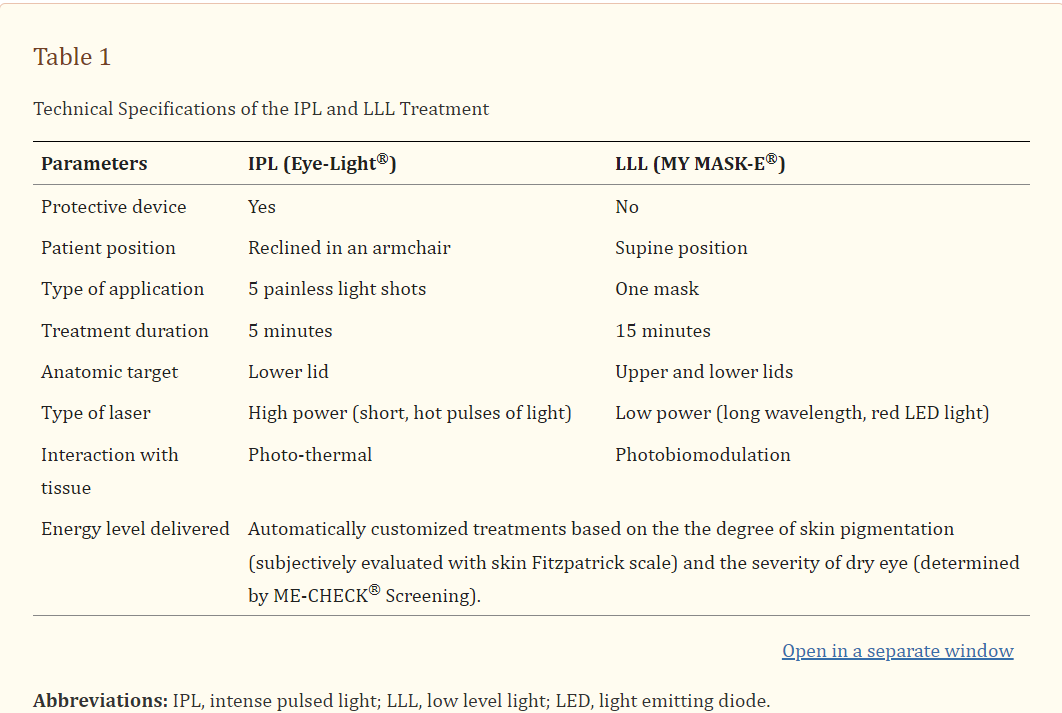
"Then, the LLLT treatment was performed applying a special mask for 15 minutes (Figure 1) (wavelength of 633 ± 10 nanometers; emission power of 100 mW per cm2; total fluence in the treated area: 110 Joules per cm2). No protective eyewear is indicated during this procedure. However, the patients were instructed to keep their eyes closed to maximize the LLLT effect on the upper and lower lids. Every session is designed to both stimulate the function of the Meibomian glands and to soften the meibum. Sessions have been performed weekly for one month." (188).
- Tear film breakup time improved in that previous study, meaning that tears lasted longer on the surface of the eye (187)
- Then, another study combining intense pulsed light and red light therapy led to the improvement of the fat layer, which helps retain moisture in the eyes (189). Here there were three treatments over three weeks. The full text of the study shows the treatment protocol in part (190):
- The problem here - again - as you can see, is that there's no independent testing of the wavelengths, the power output, and total dosage (J/cm2).
- One more study combines 600 nm and 633 nm helps to counter meibomian gland dysfunction and greatly improves quality of life (191).
- Then, another study using 600 nm and 633 nm (192). Both signs and symptoms of dry eyes are reduced. Both intense pulsed light and red light therapy were used. The intense pulsed light has a total dose of 10–16 J/cm2. The light therapy, once again, has a dose of 103 mW/cm2 and a total dose of 110 J/cm2. Just for people not believing this, here's the full quote of that treatment protocol:
"Patients were sitting or in supine position, wearing the protective opaque goggles recommended by the manufacturer. In each eye, five IPL pulses (wavelength 600 nm, 10–16j/cm2) were applied, in this order: three along the inferior orbital rim, with the device in a vertical position trying to cover all the area close to the eyewear’s edge, one behind the lateral canthus, and one along the inferior orbital rim with the device placed horizontally.
After the IPL treatment, the protective eyewear was removed and the LLLT mask was applied. It contains a series of LEDs at wavelengths of 633 ± 10 nm, with an emission power of 103 mW/cm2. During the 15 minutes of treatment, a total fluence of 110 j/cm2 is applied in the treated area. The periorbital area was treated with the patient keeping her/his eyes closed, to completely encompassing both lids." (193)
- One study once again combines intense pulsed light and red light therapy (195). Meibomian gland disease was reversed partially with a measurable improvent in 70% of eyes and a bigger one in 28% of eyes. Before treatment, tears broke up in less than 6 seconds in 85% of eyes but after treatment only in about 34% of eyes. The treatment protocol isn't fully described yet once again though (196).
- Then, lastly, on light therapy for dry eyes, there's one study using 400 - 2,000 nm polychromatic light (so the entire range). There were huge improvements, but a gel was also used in the light therapy group (197).
Then, animal studies on red light therapy for dry eyes:
- A rat study uses 680 nm, 780 nm, and 830 nm for dry eyes (170). Tear volume improved, as well as the physiological dynamics of the cornea. The light also protected to cornea damage and lowered inflammation.
- A 740 nm light mice study shows positive effects (194). Light therapy was applied once every three days. Light therapy reversed irregularities of the cornea, and improved tear volume
6) Glaucoma
Glaucoma is currently the leading cause of blindness on this planet (198; 199; 200). The condition is considered irreversible and affects 3.5% of the world's population (198). Old age is a big risk factor for glaucoma. Nevertheless, lifestyle factors play a role as well - such as excess blue light exposure (201).
The likely biological mechanism behind glaucoma is a rise in Intra-Ocular Pressure (IOP). Degeneration of the optic nerve is finally what causes the loss in vision, because of the IOP.
Here's what a recent review states about the development of glaucoma:
"In recent years, excessive light exposure has been suggested as contributing to the rise in glaucoma among the younger generation. Blue light induces mitochondrial apoptosis in retinal ganglion cells, causing optic damage; red light increases cytochrome c oxidase activity in the electron transport system, reducing inflammation and increasing antioxidant reactions to promote cell regeneration. In conclusion, the minimization of blue light exposure and the general application of red light treatment strategies are anticipated to show synergistic effects with existing treatments for retinal disease and glaucoma and should be considered a necessary prospect for the future." (201).
Only 2 human studies are available on this topic, and one is retracted, so I cannot give any specific recommendations. The overall outcomes do seem very promising though, with strong effects of near-infrared light.
Unfortunately, we don't have many red light therapy studies for glaucoma as of right now:
- One human study - that's retracted, unfortunately - did show vision improved in 70% of treated eyes with 780 nm light (202; 203). IOP also dropped by 40%. 50% of eyes had their vision restored. The transition between the cornea and sclera area with 10 J/cm2 and the eyeball with 30 J/cm2. The power output used was extremely high, at 300 mW/cm2 but there were no side effects, but, the retraction paper explicitly says these doses were calculated incorrectly (203). So, it's best to ignore this study.
- Another study uses 780 nm as well and finds reductions in ocular hypertension (glaucoma) (269). One treatment at 780 nm reduced pressure by 6 points, or 25%.
- One more in vitro study exists - but I won't treat that one because I decided not to include in vitro here (204).
7) Light-Induced Damage
Light-induced damage has many of the features of Age-related Macular Degeneration (AMD) that I considered earlier (205; 206; 207; 208). Here too, the retina or macular are damaged (205).
It's hypothesized that modern LEDs may contribute here (206). Somehow the regenerative process in the eye is impeded (207). Shorter wavelengths, like blue light, carry more energy and are thus likely more damaging (208).
Red light wavelengths especially have been proven to help, mostly in animal studies, to counteract the damaging effects of high-energy light, such as violet and blue, to the eyes (such as the retina). The red light especially (near-infrared perhaps to a lesser extent) can be used as protective pre-treatment or after exposure.
For humans, the implication here although more research is needed is that red light therapy protects against the damage of blue-dominant fluorescent and LED lighting in offices and homes.
We only have access to animal studies on this topic right now, however, but because there's lots of data I think the implications are clear.
So here are the animal studies on red light therapy for light-induced damage:
- One rat study uses 484 nm (blue) 511 nm (green), and 664 nm (red) light and compared it to traditional full-spectrum white light (209). The mixture of the three colors did better than white light, in terms of retinal damage, likely because the blue is in the high 400s.
- Red light at 670 nm protected rats' eyes, specifically the retina and surrounding tissues (210). Ingesting saffron may have similar effects.
- Exposure to 670nm protected rats' retinas against light damage (211). Other studies in rats and mice have the same outcome (212; 213; 216; 217; 218; 219; 220; 221; 222; 223; 224; 225). Photoreceptors were protected and loss of retinal tissue was slowed down. Inflammation was reduced too. Both pre-treatment and post-treatment work here. Lower doses such as 4 Jc/cm2 or 9 J/cm2 are generally used, with power outputs of around 60 mW/cm2.
- The higher the damage of light, the greater the dose of red light necessary to counter it in an animal study (214).
- 670nm and 830nm may protect neural tissue, such as after a spinal cord injury or traumatic brain injury in rats (215).
8) Myopia
About 50 different studies exist on myopia in Heiskanen's incredible chart. Myopia entails not being able to see objects far away (226; 227; 228; 229; 230; 231; 232). In myopia the eyeball grows to the wrong size, so that it's too long, so that the light doesn't focus correctly on the retina. You can't see objects that are far away, as a result.
Exposure to different colors of light may be part of what's causing myopia, as other eye problems earlier such as light-induced damage (226). Myopia often develops in childhood or young adulthood (227).
Myopia isn't purely genetic (227). Lots of screen time, for instance, is a big risk factor for developing myopia (228). Spending tons of time indoors is also a risk factor (229). Smart screen time increases myopia risk by 25%, and combined with computer time the risk goes up 77% (230). Having one myopic parent doubles your own risk, and two parents yields a 5X risk (232).
(More on this topic later, in the section about sunlight).
Spending more time outdoors is also a treatment for myopia (229; 231). Other commonly used options are:
"optical interventions such as the bifocal/progressive spectacle lenses, soft bifocal/multifocal/extended depth of focus/orthokeratology contact lenses, refractive surgery, and pharmacological treatments." (229).
Spending more than 40 minutes per day outdoors also reduces risk (232).
The low-red wavelength in particular may have very beneficial effects for countering myopia, especially when you're young. I'm talking about the 620-650nm wavelength range.
Structural changes to the eye, such as elongation of the eyeball, are reversed with that red light. Some animal studies also show that blue and UV light can be helpful in certain contexts - but we need human studies for confirmation here.
Overall, the studies don't define the power output really well, but what is important is that light is transmitted directly the pupil to deeper tissues such as the retina and choroid, to reverse myopia changes.
So let's look at the research of red light therapy for myopia right now - again, this is the best-studied topic with almost 50 studies available right now. First, human studies:
- One human study used red light therapy for myopia with night lenses (233). Children with ages of 8 - 13 were included. The elongation rate of the eye was stopped and reversed in the group with the red light therapy, but not the group with night lenses. No info was given about the red light treatment protocol though.
- Another study in children from 4 - 13 years old showed similar results (234). Results in normalizing the eyeball progressed over a 12-month period. No info is given about the red light treatment protocol.
- Then, a 650 nm study with 2 daily treatments for 3 months (235). Red light prevented and controlled myopia in children here.
- Then, 650 nm light led to less myopia in children over a 12-month period (236).
- One more study uses 650 nm twice daily for 12 months (237). The elongation of the eye was reversed in the group receiving light treatment while it worsened in the other group.
- Then there's a study using 620 nm or 875 nm (238). Here the 620 nm 77% helped shorten the eyeball in non-myopic eyes and in 41% of myopic eyes. Near-infrared at 875 nm had no effect.
- A 650 nm study used twice-per-day treatment for 6 months in children aged 7 - 14 (239). The retina and macula thickened, but light therapy seems beneficial for myopia.
- Then, one more 650 nm study led to improvements in choroidal thickness and flow (240). Axial elongation was reduced, so that the eyeball's shape improved. This study shows the development of myopia may be preventable with red light.
- One more study in just 3 children with a genetic condition that increases the risk of myopia (Sticker Syndrome) (241). Axial elongation, so the oval-shapedness of the eye, was reduced in all 6 eyes.
- Another study used different power outputs at 650 nm and shows they all help control myopia (242). There was an about 4-fold difference in power output. Too bad here the power output is measured in mW, and not simply measured as mW/cm2 with a spectrometer, which make the numbers easier to interpret.
- A study in children shows that red light improves the blood flow in the choroid (245). The thickness of the choroid also improved. Another study found a very similar outcome - the red light group improved while the control group not receiving treatment deteriorated (246).
- A 650 nm study showed a 54.1% decrease in the incidence of myopia in a 12-month treatment period (247). So light therapy prevented myopia from developing.
- Next, a study showing a decrease in eyeball elongation (248). After 12 months of treatment, about 25% of children had more normalized eyeballs, versus none in the control group. Red light at 650 nm was applied twice per day, but no power output has been measured (249).
- Then, another 650 nm study compared that treatment to spectacles (250). The red light therapy treatment was superior, with decreases in elongation of the eyeball while the spectacle treatment showed slight deterioration after 60 days.
- Another 12-month 650 nm study showed prevention of elongation in the red light treatment group versus control (251).
- Then, a 650 nm study shows less eyeball elongation and better choroid health (253).
- Another 650 nm study showed shortening of the eyeball in 25% of children, reversing myopia in children of mean age of around 10 (254).
- A 635 nm study - instead of 650 nm - shows the same outcome (255). The outcome reverses slightly though, if you quit treatment. So long-term red light exposure may be necessary to prevent myopia. Another 635 nm study has similar outcomes (260).
- Red light at 650 nm works better than atropine eye drops for preventing myopia (256). The red light outperformed the atropine 5-fold for elongation, and even bigger for refraction inside the eye.
- Then there's another twice-daily treatment study showing choroic thickness improving (257). The study took 12 months in total.
- In one more study red light outperformed a sham treatment for eyeball elongation (258).
- Another study shows that results continue to improve over a period of 2 years (259). 650 nm light was used twice per day.
- A 650 nm light at 1,600 lux was applied twice daily for three minutes outperformed spectacles (261). The same is true for lenses and red light that are compared, when lenses are worn for 6 months - red light did beter (263).
- An 830 nm study shows that it helps counter eye fatigue (262).
- Lastly, one older human study showed improvement after a single treatment (268).
Then there are animal studies on red light therapy for myopia:
- Tree shrews were exposed to 624 or 634 red light helped the animals see objects at a distance (243).
- Another study in tree shrews showed that red light had great effects, even if it was alternated with white light (244). This outcome is great because it shows that you can supplement with red light if you're overexposed to other light sources.
- A mice study using 360 - 400 nm blue light (incorrectly named "violet" in the study, showed protection against myopia progression (252).
- Bright light at 4,000 lux leads to less elongation of the eyeball than under 400 or 50 lux in guinea pigs (263).
- Research in chickens shows that blue light and UV light may help protect against myopia (264). This is thus the second animal study finding this effect, and this hasn't been investigated in humans yet. The blue and UV light even outperformed the red and white light in this study.
- Red light still helps in another rhesus monekey study, to prevent myopia (265). The monkeys were housed under fluorescent lighting. The following quote shows what I've also seen in the human studies that were extensively considered above:
"The red-light-induced alterations in refractive development were associated with reduced vitreous chamber elongation and increases in choroidal thickness. The results suggest that chromatic cues play a role in vision-dependent emmetropization in primates. Narrow-band, long-wavelength lighting prevents the axial elongation typically produced by either form deprivation or hyperopic defocus, possibly by creating direction signals normally associated with myopic defocus." (265).
- So the red light, in a normal lighting environment, helps shape the eyeball.
- Then, one more study in tree shrew (266). Blue light at 464 nm was used, as well as red light at 626 nm. Red light had similar effects as described many times before, but the eyeball returned to normal over time after exposure stopped. Flickering of light may be detrimental as well.
- Finally, there's a 624 nm and 636 nm, in the red range, in more tree shrews (267). The outcome is similar than earlier studies.
9) Optic Nerve Injury
Optic nerve damage affects the transmission of the retinal signal to the brain (268; 270; 271; 272; 273). These cells are called the retinal ganglion cells (RGCs). The problem, here, is that if these RGCs are damaged, such as because of a trauma, or oxidative stress,inflammation, or diseases such as the glaucoma I talked about earlier, you can end up with permanent vision loss (268).
Nevertheless, not all hope is lost. A recent review writes:
"After severe ON damage in optic neuropathies, promoting RGC axon regeneration is vital for restoring vision. Clearance of neuronal debris, decreased intrinsic growth capacity, and the presence of inhibitory factors have been shown to contribute to the failure of post-traumatic CNS regeneration." (268).
So RGCs can survive under the right circumstances. Generally, you'll want to keep inflammation down (270). And the good thing is we know for a fact that red light therapy is great for lowering inflammation across the body. Recently, researchers have also posited other biological mechanisms for optic nerve regeneration (272; 273).
Here, red light at around 650-670 nm seems to promote greater survival of the optic nerve and retinal cells after injury. The results are quite dramatic, although not perfectly tested in humans yet.
Arguably, some exposure of the brain and the eye area of about 4-30 J/cm2 is likely helpful and protective against optic nerve injury.
I found 9 studies on red light therapy for optic nerve injuries in total - which are only animal studies:
- A 670 nm mice study protects the RGCs against injury, by altering inflammation in part (274). The study used 4 J/cm2 for 5 days in a row.
- Another study used 670 nm at about 32 J/cm2, and that treatment prevented the damage to the optic nerve (275). The researchers here speculate that this result may also be helpful for glaucoma and Alzheimer's Disease.
- Next, 660 nm was used in hamsters (276). With treatment, 65-66% of RGCs survived, while only 45-47% survived in the control group.
- Then there's a rat study at 670 nm showing differences in short and long-term treatments:
"Short term (1 day) 670 nm light treatment is associated with reductions in reactive species at the injury site. In optic nerve vulnerable to secondary degeneration superoxide in oligodendrocytes is reduced relative to handling controls, and is associated with reduced paranode abnormalities. Long term (3 month) administration of 670 nm light preserves retinal ganglion cells vulnerable to secondary degeneration and maintains visual function, as assessed by the optokinetic nystagmus visual reflex." (277)
- Next up, another 670 nm rat study using 20 mW/cm for a whopping 30 minutes (278). Oxidative stress was lowered and the effects of nervous system injury were countered.
- Then, a study using 630 nm light (279). The light prevented rats from being affected by toxicity. The mitochondria are one of the key mechanisms of preventing that toxicity.
- Short-term exposure to 904 nm in rats prevented nerve degeneration (280). Treatment before or immediately after injury worked. Too high of a dose decreased the results, however, although the dose isn't written down carefully.
- Next, rats treated with 633 nm light (281). Exposing the damaged optic nerve of rats helped retain their health. Treatments over a period of 2 weeks were superior to 1 week.
- Lastly, there's one more 633 nm study in rabbits (282). The rabbits were exposed for 8-14 days and protected against nerve degeneration.
11) Oxygen-Induced Retinopathy
Retinopathy of Prematurity (RoP) - indicating problems with the retina because of prematurity.
On this topic, there's insufficient human evidence available to recommend treatment, although animal studies are promising.
Two animal studies explore this condition (283; 284). There, 670 nm light is protective against the effects of excess oxygen (283). The other study also uses 670 nm light protected the retina against the effects of excess oxygen once more (284).
Then, there are two human studies using 670 nm as well (285; 286). In the first study, 9 Joules / cm2 was delivered during 15 min per day, for 24-29 weeks but the study doesn't describe the outcome (285). The second study the red light boosted survival of the children that were born early although the difference wasn't statistically significant (286)
12) Retinal Blood Flow
Retinal blood flow is conerned with what the name already implies. One human study is available here, but that study doesn't just concern red light but uses polychromatic light from 600 all the way to 1,600 nm (287). Here's what the study finds:
"In 10 healthy volunteers, [red light therapy] (0.92 W, 1 : 1 duty cycle, 10 min) to both the [stellate ganglion - located near the neck] and [common carotid artery - which supply the neck and head with blood] significantly increased peak systolic blood velocity in the ophthalmic artery (p<0.001, each) and central retinal artery (p<0.001, each) without changes in vessel resistance. Irradiation to the [common carotid artery] produced a stronger effect than that to the [stellate ganglion] in the ophthalmic artery (p=0.007) and central retinal artery (p=0.031)." (287).
This is good news, as irradiating the neck and head likely has an effect on the retina! So for people who cannot stand any light exposure to the eyes, treatment of the neck and head may have systemic effects that affect eye health in the end as well.
13) Retinitis Pigmentosa
Retinitis pigmentosa (RP) is a hereditary condition affecting the retina (288; 289; 290; 291; 292). RP leads to progressive vision loss and is hard to treat (288). RP often affects both eyes (290).
As the name already implies, RP is characterized by changes of eye color - here the retina turns purple and red (288). Two different photorecpetors in the eye, the "rods" and "cones", are eventually destroyed (289). Rods help you see in dim lighting, in black and white, and make up 95% of photoreceptors (289). Cones help you see color and require more light for you the photons to be converted into an impulse the brain can deal with (289)
Gene therapy is often applied for severe RP and nutrition for its early stages (288; 291). Often, there already are vision problems from childhood (289). The condition is very hard to treat, though (289). Often, because the macula stays intact, people will be able to see something still (290). About 1 in 5,000 people are affected by RP worldwide (290).
Huge improvements are possible in many retinitis pigmentosa studies with light therapy. However, more human research is needed for confirmation as study quality is a bit low.
Generally though, it seems that high-powered red and near-infrared light, for short treatment periods, can have great effect on RP.
Red light therapy for retinitis pigmentosa has an effect as well - I'll first consider human studies:
- One human study uses 633 nm for 3 minutes per day, either at 25 mW/cm2 or 100 mW/cm2 (292; 293). After 8 weeks of treatment, participants recovered 5.4 letters of vision, when viewed at a distance. The 100 mW/cm2 group improved more than the 25 mW/cm2 group. It's hypothesized here that the rods specifically are affected by the red light therapy in this study.
- Secondly, a 780 nm study that also shows "significant improvement" - although the study was a case of just 1 participant (294). Nine sessions of red light therapy every other day, both regular visual acuity during the daytime, night vision, and quality of life improved dramatically.
- Then, one more study using 633 nm in humans with RP with the treatment described as "highly effective" (296). Study participants' vision acuity improved after the therapy for a period of three years. The dose is not described well in the study, and implies only 1 mW/cm2 was used for 3 minutes per treatment, once every 2 days.
- Lastly, one more human study shows a huge dose of 333 mW/cm2 at 780 nm, applied for 40 seconds, helps improve vision and maintain that improvement (297). Treatment ensued twice weekly for two weeks. Five years after treatment, the results had reversed, but after four new treatments, the patient regained their vision. These results are crazy because treatment in a total of 3 minutes (over 4 sessions) can lead to huge improvements in RP.
Then, animal studies no red light therapy for retinitis pigmentosa - there's only one study:
- Here rats were treated with 830 nm, at 25 mW/cm2, for 3 minutes (295). The rats that were treated with light had better metabolism in their retina, retained their retina structure better, and had better retinal function. Oxidative stress in the light treated group was lower.
Assorted Eye Conditions
Some other eye condtions have very poor research backing them in terms of light therapy, such as Amblyopia or Chalazia, and I've decided not to include them to prevent people from treating it if there's only a single study available. Apologies!
How to Use Red Light Therapy for Eyes: Expert Tips
So how should you use red light therayp for the esy? Given all the evidence I've listed above, it's probably best to expose your eyes to 40 - 60 mW/cm2 for 3 minutes per day. You can split these up into two sessions, if necessary.
Some studies use higher power outputs but I've consistently seen good outcomes with the 40 - 60 mW/cm. And, the light needs to enter the pupil, so you cannot wear protective glasses.
So, if you want maximum safety with maximum benefit, you'll use your red light therapy panel for 3 minutes and stand in front of it so the light can enter the eyes. Make sure the distance is sufficient so that you're getting a 40 - 60 mW/cm2 dose. And then, when your 90 - 180 second session is over, wear the protective eyeglasses.
Theoretically, you could also use red only, as there are more studies on red light than near-infrared. But as the near-infrared light studies are also positive, I'd probably opt for using them both, especially as the near-infrared light will penetrate deeper into the eyes and therefore affect more of the retina and macula.
Red Light Therapy For The Eyes Safety
A few studies have been specifically written about red light therapy for eyes safety (296; 297; 298). Sometimes power outputs are too high and therefore become damaging to eye health.
High-powered incandescents and halogens can potentially have negative effects, for instance (296). In rat studies, 500 mW/cm2 can be damaging to the retina for some (297). And when 7,200 mW/cm2 is used, it can also be problematic (298).
All of these conclusions are somewhat common sense. Just use normal power output levels and your eye health should be fine. So, many of the studies used 40 or 60 mW/cm2, for several minutes. Often, that's the same power output you'd get by standing directly in front of a panel for several minutes.
NASA states that the sun can have a power output of up to 137 mW/cm2 - when it's directly overhead (299). That sun also emits the highly energetic ultraviolet A and B (UVB and UVB), as well as the far more heating mid or medium-infrared (MIR) and far-infrared (FIR). Red light therapy products never emit any UVA, UVB, MIR, and FIR, and should thus be safe to use for the eyes at normal power intensities.
Red Light Therapy For Vision: Assorted Topics Such As Nutrition, Sunlight, And More
Lastly, I want to talk about a few assorted topics that I've not covered before. I briefly want to state something about these topics because I don't want people to get away with the idea that you just need red light therapy to maintain eye health.
As almost always, health is a multi-factorial endeavor. Just like your blood sugar levels aren't just affected by light exposure, but also by exercise, by nutrition, by sleep quality, by the air you breathe and how you breathe it, red light therapy for vision alone isn't enough to guarantee the optimal outcome
So let's very briefly talk about these topics:
Sunlight And Eye Health?
I'll probably write a blog post about this topic if it interests people. I won't go into much detail here.
Nevertheless, I will say that sensible sunlight exposure - without getting sunburns or spending hours per day in the bright sun, is likely very protective for good eye health. Spending time outdoors is highly protective against eye diseases in general, although it's more complicated than that (300; 301; 302; 303; 304).
Indoor air pollution may also play a role and increase risk - if you don't filter the air indoor pollutant levels can get 10X as high as outdoor levels (301). But, the details of this topic will be discussed in a future blog post.
For now, especially given that sunlight exposure has been part of human history forever - except for the last 150-200 years - reasonable sunlight exposure without sunglasses is recommended if you don't overdo it. But, that's the same for exercise, for dieting, for sauna use, and so forth - there's a golden mean.
Nutrients To Support Eye Health
In this section too, I don't want to get into too much detail, except for telling you that your diet has a huge influence on eye health. Here are a few simple examples:
- Vitamin D levels are at the very least strongly associated with eye health, and likely causal (302; 303; 304; 305; 306; 307). Both sunlight and vitamin D levels play a role in myopia (302). Vitamin D levels are associated with other diseases such as dry eye syndrome, Age-related Macular Degeneration (AMD), diabetic retinopathy, glaucoma, and a whole host of others (303; 306). Reversing vitamin D deficiency through supplementation can alleviate some eye conditions such as dry eyes (304; 305).
- Carotenes, like beta-carotene, lutein and zeaxanthin (308; 309; 310; 311; 312). Here's what a recent review states about carotenoids. Carotenoids:
"have distinct physiological and pathophysiological functions ranging from fetal development to adult homeostasis. β-carotene is a precursor of vitamin A that essentially functions in many biological processes including vision. The human macula lutea and eye lens are rich in lutein, zeaxanthin, and meso-zeaxanthin, collectively known as macular xanthophylls, which help maintain eye health and prevent ophthalmic diseases. Ocular carotenoids absorb light from the visible region (400-500 nm wavelength), enabling them to protect the retina and lens from potential photochemical damage induced by light exposure. These natural antioxidants also aid in quenching free radicals produced by complex physiological reactions and, consequently, protect the eye from oxidative stress, apoptosis, mitochondrial dysfunction, and inflammation." (308)
- Animal foods, fruits, and vegetables are excellent carotenoid sources. Caretonoids like lutein - which is most available from eggs and green leafy vegetables - are directly tied to the risk of developing eye diseases (309; 310). AMD is one example here (312). Carotenes also improve night vision (311).
- Magnesium deficiency is strongly linked to eye conditions (313; 314; 315; 316; 317). A recent review states:
"Recent studies highlighted the association of hypomagnesemia and, thereby, supplementation of Mg2+ in the management of eye diseases. Glaucoma, senile cataract and diabetic retinopathy were associated with low level of extracellular Mg2+. The neurovascular protective effects of Mg2+ mediated through activation of endothelial nitric oxide synthase and inhibition of endothelin-1 eventually result in vasodilatation of retinal vessels." (313).
- Magnesium is omnipresent in modern society and alters fundamental physiology in the eye (314). Having low magnesium compared to sodium may alter blood flow dyanmics in the ey, for instance (314). Magnesium deficiency may also cause oxidative stress in the eye (314). Magnesium deficiency also changes the structure of the retina and optic nerve (315).
- Many of the nutritional deficiencies, such as carotenoid and magnesium, can increase the odds of eye or retina conditions by 20% or so, individually (316; 317).
- Adequate zinc affects the risk and progression of some eye diseases (318; 319; 320; 321; 322).
Overall the message is simple: eat a balanced, healthy diet. Cut out processed and hyperpalatable foods, and soda, etc. But again, don't focus on diet only, like you shouldn't focus on red light only, as other factors such as sleep quality and many other matter as well (323; 324; 325; 326; 327; 328; 329; 330).
Frequently Asked Questions
Finally, let me answer a few frequently asked questions - you should be able to guess the answer to these questions by now, if you're read my blog post:
Is Red Light Therapy Safe For Eyes?
Red light therapy is absolutely safe for eyes, if you use the correct dose that I've described above. If you underdo it (so you don't expose your eyes to any light) or overdo it (you expose your eyes to excess light), you'll get suboptimal results.
What Are the Benefits Of Red Light Therapy for Eye Health?
Red light therapy can normalize eye health for many people with eye conditions, ranging from myopia to diabetic retinopathy to dry eyes. Unfortunately there's not much research on improving eyesight and health in already healthy eyes...
How Often Should I Use Red Light Therapy For My Eyes?
A treatment frequency of 3-4 times per week, for 3 minutes at a time at 40 - 60 mW/cm2, is probably optimal. You can use a tabletop panel or n a handheld panel as a cheaper treatment option, if you want to use red light therapy for vision purposes only.
If you want to treat your entire body, consider getting a wall panel or bigger. The wall panels can be used for eye health, if and only if you get the correct power output into your eyes for the correct amount of time.
Do You Need to Cover Eyes While Using Red Light Therapy?
If you always cover your eyes during red light therapy, the benefits will diminish because the light isn't reaching all the tissues of your eyes.
Why Do I See Green Colors After Treatment?
If you're exposed to lots of red and near-infrared light, some people's perception changes for a short while after treatment. That's normal and you should return to your normal color vision quickly after a treatment!
What Are Potential Side Effects For Red Light Therapy For Vision And Eye Health?
With excess treatment time or power output, you may get retinal or macular damage over time. So, I don't recommend exceeding the treatment guidelines that I've laid out earlier in this blog post.
If you really want to treat your eyes with red light therapy, I do recommend doing so under medical supervision if you want to remove any risk of side effects.
To fully avoid any side effects, I still recommend seeking out professional medical help - so nothing in this blog post is medical advice. Also, if you've got surgery for your eyes, I'm not willing to take the risk of giving any recommendations here.
Conclusion: Red Light Therapy For The Eyes Can Be A Game-Changer
Hopefully you're getting tons of benefit from my analysis and summary of all available evidence on red light therapy for the eyes! I'll likely update this blog post in the future, but it will be a lot of work.
If you liked this blog post, please support my work by sharing this blog on social media! You may likely help lots of loved ones with this blog post as well - and keep in mind I've given you all of this data at no cost at all!
Want More Deep Dives On Light Therapy? Join My Advanced Science Course Below:

That course teaches you:
- The science on many different goals, whether diseases or maximizing performance
- Best device selection based on scientific criteria
- Maximizing your benefits for different goals, whether gym performance, sleep quality, using UV light, or more!
Join for free HERE!
Found This Interesting? Then You Might Like:
- Start Here - Light Therapy 101 & Buyers Guide
- Discount Codes, Deals & Recommendations - Red Light Therapy
- Red Light Therapy For Upgrading Your Brain Health
- Does Vielight Work? A Close Look At The Spectacular Vielight Science
- Red Light Therapy For Weight Loss: The Science Of Supercharging Fat Loss
- Red Light Therapy For Sleep: The Science of Sweet Dreams
- The Effectiveness Of Light Therapy For Sleep Disorders
This is a post by Bart Wolbers of Lighttherapyinsiders. Bart finished degrees in Physical Therapy (B), Philosophy (BA and MA), Philosophy of Science and Technology (MS - with distinction), and Clinical Health Science (MS), has had training in functional medicine, and is currently chief science writer.